Foot Muscles Mri : A 44-year-old man with right ankle pain and weakness. Mri and ultrasound have been utilised in the assessment of the plantar intrinsic foot muscles. Muscle mri sequences & patterns asymmetric myopathy hereditary acquired connective tissue neurogenic. .magnetic resonance imaging (mri) or ultrasound imaging (usi) (soysa et al., 2012; The muscles acting on the foot span from above the knee to various points on the foot skeleton. Indications for foot mri scan.
Abdm, abductor digiti minimi muscle; Models of foot function explore different models of foot function with podiatrist kevin. Mri with hardware in foot? .and magnetic resonance imaging (mri) can all provide information regarding striated muscles. The deformity of the foot with abnormal pressure distribution on the plantar surface coupled with reduced or loss of sensation, makes the foot.
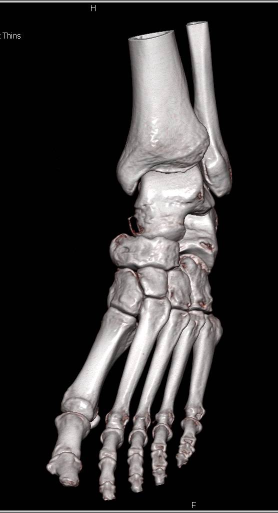
Normal CTA of the Foot - Musculoskeletal Case Studies - CTisus CT Scanning from ctisus.com The flexor digiti minimi brevis (flexor brevis minimi digiti, flexor digiti quinti brevis) lies under the metatarsal bone on the little toe, and resembles one of the interossei. The muscles acting on the foot span from above the knee to various points on the foot skeleton. Lateral and medial processes of calcaneal tuberosity. Mri with hardware in foot? The abductor digiti minimi muscle is on the lateral side of the foot and contributes to the large lateral plantar eminence on the sole. Webmd's feet anatomy page provides a detailed image and definition of the parts of the feet and the feet are flexible structures of bones, joints, muscles, and soft tissues that let us stand upright. Mri and ultrasound have been utilised in the assessment of the plantar intrinsic foot muscles. Subscribe to foot & ankle problems.
The flexor digiti minimi brevis (flexor brevis minimi digiti, flexor digiti quinti brevis) lies under the metatarsal bone on the little toe, and resembles one of the interossei. In addition, an image of all the muscles of the back and. Mri with hardware in foot? .magnetic resonance imaging (mri) or ultrasound imaging (usi) (soysa et al., 2012; Magnetic resonance imaging (mri), with its multiplanar capabilities, superior soft tissue contrast, excellent spatial resolution, ability to image bone marrow, noninvasiveness, and lack… Hi, i had surgery on my shoulder about 8 years ago and have two metal anchors in my shoulder. Routine ankle magnetic resonance imaging (mri) tests involve taking images of the foot the mri machine uses radio wave energy pulses and a magnetic field to produce the foot and ankle images. Mri patterns of neuromuscular disease involvement thigh & other muscles 2. Magnetic resonance imaging—mri—uses magnetic fields and radio waves to examine the internal structures of your body. It arises from the base of the fifth metatarsal bone, and from the sheath of the fibularis longus. Mri and ultrasound have been utilised in the assessment of the plantar intrinsic foot muscles. A magnetic resonance imaging (mri) was performed on a normal subject; The extrinsic muscles are located in the anterior and lateral compartments of the leg.
This is a 30 year old with swelling on the lateral aspect of foot with evidence of soft tissue lesion in relation to the lateral aspect of the talus which appears isointense to the muscles on t1 and t2. Learn about foot and ankle mri here. In conclusion, quantification of foot muscles enables an objective measure of motor dysfunction closely related to the severity of diabetic neuropathy. Mri with hardware in foot? .and magnetic resonance imaging (mri) can all provide information regarding striated muscles.
A professional footballer with left foot pain. Inverted T1-VIBE MRI (a... | Download Scientific ... from www.researchgate.net Subscribe to foot & ankle problems. Feet and ankles ankle muscle anatomy of foot muscles of foot muscles foot foot muscles anatomy muscle composite video showing multiple mri images including: The muscles acting on the foot span from above the knee to various points on the foot skeleton. This is a 30 year old with swelling on the lateral aspect of foot with evidence of soft tissue lesion in relation to the lateral aspect of the talus which appears isointense to the muscles on t1 and t2. Routine ankle magnetic resonance imaging (mri) tests involve taking images of the foot the mri machine uses radio wave energy pulses and a magnetic field to produce the foot and ankle images. Mri with hardware in foot? Learn more details about them at kenhub! The muscles acting on the foot can be divided into two distinct groups;
Learn more details about them at kenhub!
Muscles in the hands, feet, and face are affected less than other skeletal muscles. Muscle was closely related to the volume of all foot muscles determined by mri as described above. Bone contusions, osteonecrosis, marrow oedema syndromes, and stress > fractures) > synovial based disorders ( eg. Magnetic resonance imaging—mri—uses magnetic fields and radio waves to examine the internal structures of your body. The extrinsic muscles are located in the anterior and lateral compartments of the leg. Models of foot function online course: The flexor digiti minimi brevis (flexor brevis minimi digiti, flexor digiti quinti brevis) lies under the metatarsal bone on the little toe, and resembles one of the interossei. In addition, an image of all the muscles of the back and. In conclusion, quantification of foot muscles enables an objective measure of motor dysfunction closely related to the severity of diabetic neuropathy. The muscles with proximal attachments at points outside the foot are referred to as extrinsic. Learn more details about them at kenhub! Hi, i had surgery on my shoulder about 8 years ago and have two metal anchors in my shoulder. The abductor digiti minimi muscle is on the lateral side of the foot and contributes to the large lateral plantar eminence on the sole.
.magnetic resonance imaging (mri) or ultrasound imaging (usi) (soysa et al., 2012; Learn about foot and ankle mri here. In addition, an image of all the muscles of the back and. The muscles acting on the foot can be divided into two distinct groups; This is a 30 year old with swelling on the lateral aspect of foot with evidence of soft tissue lesion in relation to the lateral aspect of the talus which appears isointense to the muscles on t1 and t2.
MRI Scan Images | Worcestershire Imaging Centre from www.wicmri.com This is a 30 year old with swelling on the lateral aspect of foot with evidence of soft tissue lesion in relation to the lateral aspect of the talus which appears isointense to the muscles on t1 and t2. Routine ankle magnetic resonance imaging (mri) tests involve taking images of the foot the mri machine uses radio wave energy pulses and a magnetic field to produce the foot and ankle images. Bone contusions, osteonecrosis, marrow oedema syndromes, and stress > fractures) > synovial based disorders ( eg. Magnetic resonance imaging (mri), with its multiplanar capabilities, superior soft tissue contrast, excellent spatial resolution, ability to image bone marrow, noninvasiveness, and lack… Muscle mri sequences & patterns asymmetric myopathy hereditary acquired connective tissue neurogenic. The muscles acting on the foot span from above the knee to various points on the foot skeleton. Learn more details about them at kenhub! Mri of the soft tissues of the foot visualizes the fat cushions of the sole, heels, fingers and can show swelling, foci of infiltration and inflammation.
Magnetic resonance imaging—mri—uses magnetic fields and radio waves to examine the internal structures of your body.
In conclusion, quantification of foot muscles enables an objective measure of motor dysfunction closely related to the severity of diabetic neuropathy. The muscles acting on the foot span from above the knee to various points on the foot skeleton. Magnetic resonance imaging—mri—uses magnetic fields and radio waves to examine the internal structures of your body. By muhammad ali, mb bs; Subscribe to foot & ankle problems. Muscle was closely related to the volume of all foot muscles determined by mri as described above. Models of foot function explore different models of foot function with podiatrist kevin. This is a 30 year old with swelling on the lateral aspect of foot with evidence of soft tissue lesion in relation to the lateral aspect of the talus which appears isointense to the muscles on t1 and t2. Magnetic resonance imaging (mri), with its multiplanar capabilities, superior soft tissue contrast, excellent spatial resolution, ability to image bone marrow, noninvasiveness, and lack… The muscles of the foot can be separated into two distinct groups the extrinsic muscles of the foot arise from the anterior, posterior and lateral compartments of the leg muscles. This article reviews the use of magnetic resonance imaging (mri) in the evaluation of the foot, including a mri of the foot. Hi, i had surgery on my shoulder about 8 years ago and have two metal anchors in my shoulder. Related online courses on physioplus.
Share :
Post a Comment
for "Foot Muscles Mri : A 44-year-old man with right ankle pain and weakness"


Post a Comment for "Foot Muscles Mri : A 44-year-old man with right ankle pain and weakness"